
External urethral sphincter: function
The external urethral sphincter for the pelvic health clinician: what it closes, how a cough moves it, and how to train it.

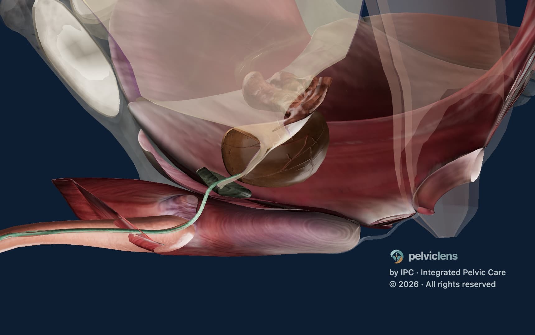
The short answer. The external urethral sphincter is the striated muscle loop that closes the urethra below the bladder neck (the membranous urethra in men, the mid-urethra in women) and relaxes for voiding. A voluntary squeeze displaces the mid-urethra dorsally, and a cough recruits the sphincter in its own reflex pattern [1] [2]. After radical prostatectomy it is the main closure left.
Key takeaways
- Of the pelvic floor's three layers, only the levator ani and the striated urethral sphincter make urethral pressure; the bulbospongiosus compresses the bulb of the penis and adds none.
- A voluntary squeeze and a cough recruit the sphincter in different patterns, and a stress leak tests the cough's reflex pattern, not the squeeze your patient performs on request.
- A submaximal contraction lifts the bladder neck almost as far as a maximal one and can be held far longer. Continence control places that gentle squeeze right before the cough, the lift or the stand.
- In women, urethral closure pressure falls with age while support holds steady, so a leak in later life is often a sphincter problem. The operations that improve support do not typically change closure pressure.
Most references give the external urethral sphincter one line: a ring of striated muscle under voluntary control, strengthened by Kegel exercises. Ask a healthy man to cough under transperineal ultrasound, and the screen shows what that line leaves out. The levator ani lengthens while the sphincter shortens and displaces the mid-urethra dorsally, though nobody asked for a pelvic floor contraction [1]. The squeeze your patient performs on request and the closure that holds a cough are two patterns of one muscle, and it is the second that a leak tests.
What is the function of the external urethral sphincter?
The contraction you see when you ask for a squeeze is not the one that holds a cough. IPC's teaching describes a voluntary contraction layer by layer. The levator ani displaces the urethrovesical junction cranially, the striated urethral sphincter in the middle layer displaces the mid-urethra dorsally, and the bulbospongiosus compresses the bulb of the penis. Of the three, only the levator ani and the sphincter make urethral pressure.
A cough recruits the same muscles in another pattern. The levator ani lengthens and the urethrovesical junction displaces dorsally, while the sphincter still displaces the mid-urethra dorsally and the bulbospongiosus still compresses the bulb. In neurologically normal men, a cough raised urethral closure pressure at the distal posterior urethra, with urethral pressure rising about two and a half times as much as rectal pressure [2]. In men with neurological lesions the cough response and the voluntary squeeze failed independently of each other, as expected of a reflex and a voluntary response [2].
Between coughs the sphincter holds tone. On EMG it is tonically active through storage and silent once voiding begins [3]. Its firing rises as the bladder fills, driven in part by the pudendal guarding reflex [4]. A radical prostatectomy resects the internal sphincter with the prostate, so the striated sphincter in the middle layer and the levator ani behind it are the closure that remains. A cough that pushes bladder pressure above the closure pressure they generate produces the leak of intrinsic sphincter deficiency.
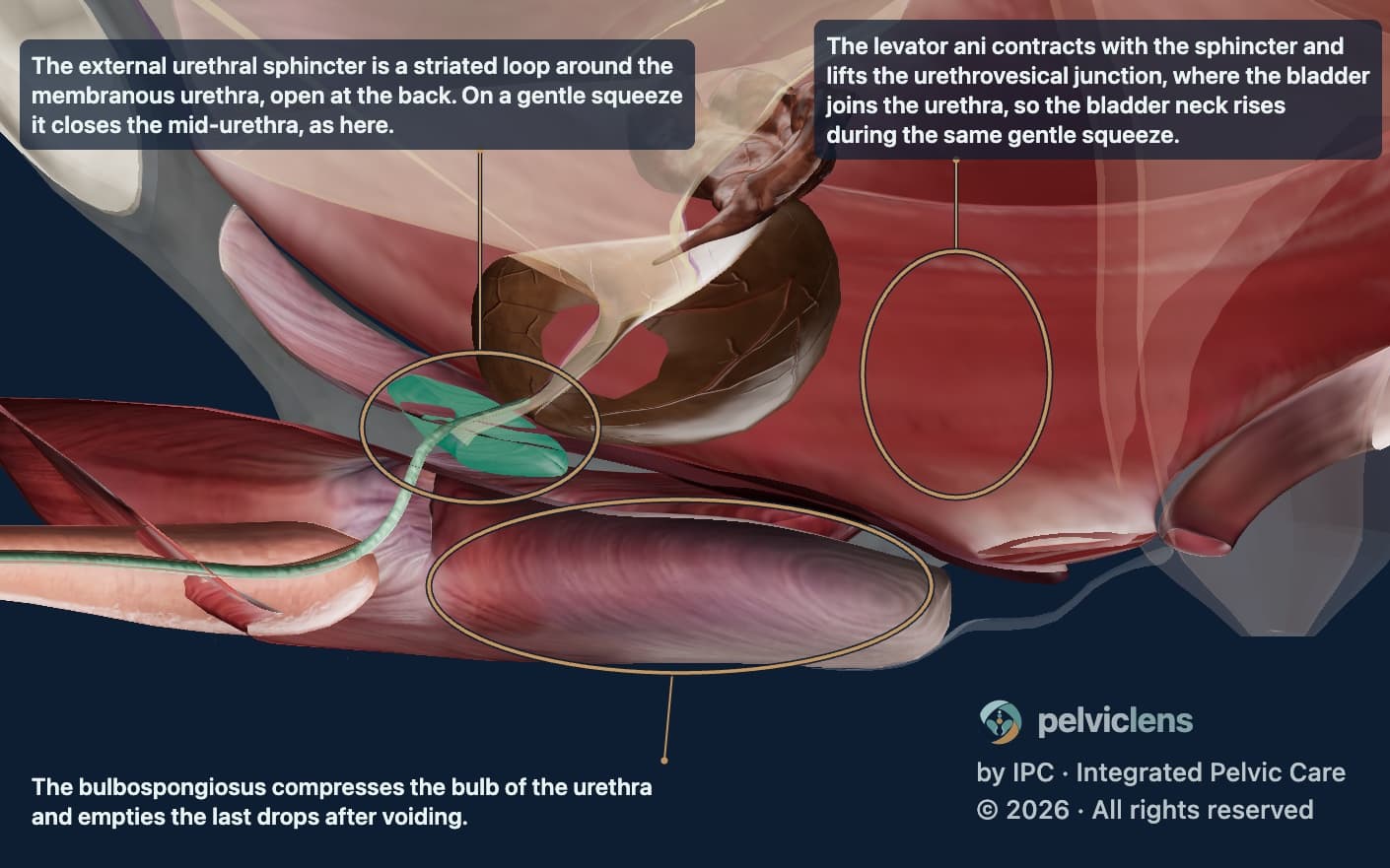
This still, from the formula chapter of the Pelvic Lens incontinence model, shows intact male anatomy with the prostate in place during a held gentle squeeze. At that moment the model's urethral closure pressure is 129 cmH2O, 74 at rest plus 55 from the squeeze, while its bladder pressure stays flat. The three circles mark the striated urethral sphincter closing the membranous urethra, the levator ani lifting the urethrovesical junction above it, and the bulbospongiosus compressing the bulb of the penis below it. The full animation plays on pelviclens.com.
External urethral sphincter anatomy: the middle layer
The ring in the textbooks is the wrong picture of this muscle. The teaching describes the striated urethral sphincter as an omega-shaped loop, open posteriorly, with its origin and insertion both at the central perineal tendon. So the loop is anchored behind the urethra rather than closed around it [1]. Reconstructions of fetal and adult pelves show that the loop has no direct bony attachment [5]. In the female pelves of that series a tendinous connection anchored the sphincter to the levator ani, an attachment the male sphincter lacked. Where that attachment exists, the two muscles contracting together bend the mid-urethra and close its lumen [5].
The fibre types correct the textbook as well. The classic histology found only slow-twitch, type I fibres, adapted to hold tone over long periods [6]. A later series of male specimens measured about 30 percent fast-twitch fibres, mostly the fatigue-resistant type 2A. The sphincter's fibres were also small, about a third of the levator ani's in mean cross-sectional area [7]. The teaching reads that mix as two jobs: the slow fibres hold tone through storage, and the fast, fatigue-resistant ones close the urethra quickly when a cough arrives.
External urethral sphincter after prostatectomy
Every account of post-prostatectomy incontinence lists what the operation removes; your work starts from what it leaves. The internal sphincter is resected with the gland, and the bladder neck is resected too or, where it is preserved, divided and reconstructed. The bladder is then anastomosed to the membranous urethra, whose striated loop the operation leaves in place.
What returns over the months is the sphincter's function, not its length. In one series, continence rose from 18 percent at six weeks to 82 percent at six months. Over the same months the functional sphincter length, 50 mm before surgery, stayed at 24 to 25 mm in continent and incontinent men alike [8]. What recovered was posterior urethral sensation and cough pressure transmission, the share of a cough's pressure rise that reaches the urethra [8]. The patient guide to post-prostatectomy recovery, which lays out that curve with a bladder diary to track it, is the page to send your patient home with.
In a second series, continent men also used what was left differently. On a maximal contraction they shortened the sphincter, the puborectalis and the bulbospongiosus further than the men who leaked. On a cough their bladder neck descended less and their sphincter displaced further [9]. That way of using the muscles, not the length of sphincter left, is what training works on.
How the external urethral sphincter is trained
A harder squeeze does not hold a cough better. A maximal contraction raises bladder pressure along with urethral pressure, so your patient can leak during the exercise itself. A submaximal one adds closure without pushing on the bladder. The teaching calls the training built on that difference continence control: targeted storage strategies used right before and during the activities likely to trigger a leak. Each strategy is tested on the same cough, in the order of the Mechanical Zones. Your patient sits tall (MZ I, the Spinal Zone), breathes out with the lower abdomen drawn in (MZ II, the Abdominal Zone), and then squeezes gently (MZ III, the Pelvic Zone).
When your patient is a man, the grade of the squeeze is visible. A light contraction moves the penis and leaves the testicles still: that is the bulbospongiosus working alone. When the testicles begin to rise, the middle layer, where the sphincter sits, has joined in, and that is the level to hold, short of a maximal effort.
For the gentle squeeze, the teaching cites a study of women with and without stress urinary incontinence. There a submaximal contraction lifted the bladder neck 4 mm, within a millimetre of a maximal one, and was held for 33 seconds against 12 [10]. The maximal effort pushed the bladder neck down in more than a quarter of the women, which the submaximal one never did. It also roughly doubled the rise in abdominal pressure [10].
Train endurance, a sustained submaximal hold for the tone the sphincter keeps through storage, and reaction, a quick closure placed right before a cough, as separate goals. None of this training starts while a urethral catheter is in place. A catheter cannot be compressed, so a sphincter squeezed against it learns to close only as far as the tube allows. Once the tube is out, the urethral walls do not meet.
When it does not relax
The same muscle has to relax for the stream to flow, and the teaching examines a non-relaxing floor layer by layer. A levator ani that does not relax holds the bladder neck high, so the stream is hard to start and the bladder does not empty completely. A sphincter that does not relax compresses the mid-urethra and weakens the stream. A bulbospongiosus that does not relax compresses the bulb and leaves a post-micturition dribble. Which layer fails to relax decides the treatment. In 15 women with dysfunctional voiding or unexplained urinary retention, EMG found the pelvic floor active during voiding in 11 and the sphincter in 4. Biofeedback training helped the first group but not the second [11].
Assessing the external urethral sphincter in clinic
Your patient can hold a five-out-of-five squeeze against your finger and still leak on the first cough of the IPC stress cup test. On a voluntary contraction the levator ani lifts everything under your finger, and the mid-urethra moves out of reach. An EMG recording from a single site in the levator ani may not represent the external urethral sphincter either [6].
The teaching's test is a slow cough with your finger at the mid-urethra. As the cough builds, the levator ani lengthens and descends, and then the reflex contraction of the sphincter taps your fingertip. That tap is the only sign of the sphincter working that a physical examination gives. Even so, a cough on request raises abdominal pressure less than an evoked, reflex cough, so it may underestimate what the continence mechanism has to hold [1]. The cup test then records the result for each trigger, as drips or a jet, before and after each strategy.
Transperineal ultrasound of a voluntary contraction shows two strategies among continent men, some moving the urethra mainly with the sphincter and others mainly with the levator ani [12]. If your patient's strategy was sphincter-led before surgery, he may have to compensate with the levator ani afterwards, a pattern he has never relied on. Choose a cue that teaches him the levator-led strategy. All of this assessment sits inside the stress urinary incontinence workup. There the bladder diary reads Fluid Imbalance, Storage Impairment and Voiding Impairment, the first three of the 4Is, before the fourth, Incontinence, is attributed to the sphincter.
The external urethral sphincter in women
Your older patient who leaks on a cough is usually examined for support, and her support is often the part that has not changed. Her external urethral sphincter is one continuous striated muscle with three parts: the sphincter urethrae, the compressor urethrae and the urethrovaginal sphincter [13]. In nulliparous women aged 21 to 70, maximal urethral closure pressure fell by about 15 cmH2O a decade while pelvic organ support, urethral support and levator function held steady [14]. In a case-control study, closure pressure separated women with stress incontinence from matched continent women with an effect size more than twice that of any measure of support. Levator strength did not differ between the groups [15]. Her leak is often a story of falling closure pressure rather than failing support. The widely used operations improve urethral support and usually succeed, yet they do not typically change urethral closure pressure [15]. In my clinic, her first treatment is the same one I teach after a prostatectomy: a gentle squeeze placed right before the cough.
What to say to your patient
- With the model open: "This loop is your sphincter, the muscle that holds pee in. A small squeeze closes it."
- When you teach the cough: "Sit tall. Breathe out. Squeeze a little, then cough."
- When the stream is weak: "Let your pelvic floor go soft before you start. Once the pee flows, do not push or squeeze."
- When her support tests normal: "The tissue holding up your bladder and pee tube is fine. The muscle around your pee tube closes less tightly now. We train it to close at the right moment."
Frequently asked questions
Can you control the external urethral sphincter?
Yes, through the pudendal nerve: it is striated muscle, and a voluntary squeeze closes it on demand. A cough also recruits it, reflexly and in a pattern of its own, and that reflex closure is the one a leak tests. Training therefore times a gentle voluntary squeeze to land before the cough.
What happens when the external urethral sphincter is damaged?
Closure pressure falls, and urine leaks whenever a cough, a lift or a stand pushes bladder pressure above it. A partial loss gives stress leakage at a low effort, and a severe one can give continuous leakage, as after some prostatectomies. Training comes first and, after prostatectomy, can hasten the return of continence. Under the AUA/SUFU guideline, surgery such as an artificial urinary sphincter is considered when the leak persists at one year or is severe at six months.
How to strengthen the external urethral sphincter?
Train timing and endurance rather than maximal force. A submaximal contraction lifts the bladder neck nearly as far as a maximal one and can be held far longer, so teach it gently, place it right before the cough or the stand and hold it through. Train a sustained hold and a quick reaction separately, never with a catheter in.
What type of muscle is the external urethral sphincter?
It is striated, skeletal muscle under somatic control through the pudendal nerve. Its small fibres are mostly slow-twitch, built to hold tone all day, and nearly a third are fast-twitch, mainly fatigue-resistant type 2A, for the quick closure of a cough. It has no muscle spindles and no direct bony attachment.
References
- Dynamics of male pelvic floor muscle contraction observed with transperineal ultrasound imaging differ between voluntary and evoked coughs. Journal of Applied Physiology, 2014.
- Urethral pressure response to cough and voluntary perineal contraction in men without previous pelvic surgery. Neurourology and Urodynamics, 1999.
- What do we really know about the external urethral sphincter?. Continence, 2024.
- Neural control of the lower urinary tract. Comprehensive Physiology, 2015.
- The anatomical components of urinary continence. European Urology, 2009.
- A comparative study of the human external sphincter and periurethral levator ani muscles. British Journal of Urology, 1981.
- Striated muscle fiber compositions of human male urethral rhabdosphincter and levator ani. Journal of Urology, 2006.
- Effect of radical prostatectomy on sensory threshold and pressure transmission. Journal of Urology, 2000.
- Comparison of dynamic features of pelvic floor muscle contraction between men with and without incontinence after prostatectomy and men with no history of prostate cancer. Neurourology and Urodynamics, 2020.
- Submaximal pelvic floor muscle contractions: similar bladder-neck elevation, longer duration, less intra-abdominal pressure. International Urogynecology Journal, 2018.
- Dysfunctional voiding in women: which muscles are responsible?. British Journal of Urology, 1998.
- Novel insight into the dynamics of male pelvic floor contractions through transperineal ultrasound imaging. Journal of Urology, 2012.
- The striated urogenital sphincter muscle in the female. Anatomical Record, 1983.
- Effects of aging on lower urinary tract and pelvic floor function in nulliparous women. Obstetrics and Gynecology, 2007.
- Stress urinary incontinence: relative importance of urethral support and urethral closure pressure. Journal of Urology, 2008.
Related articles
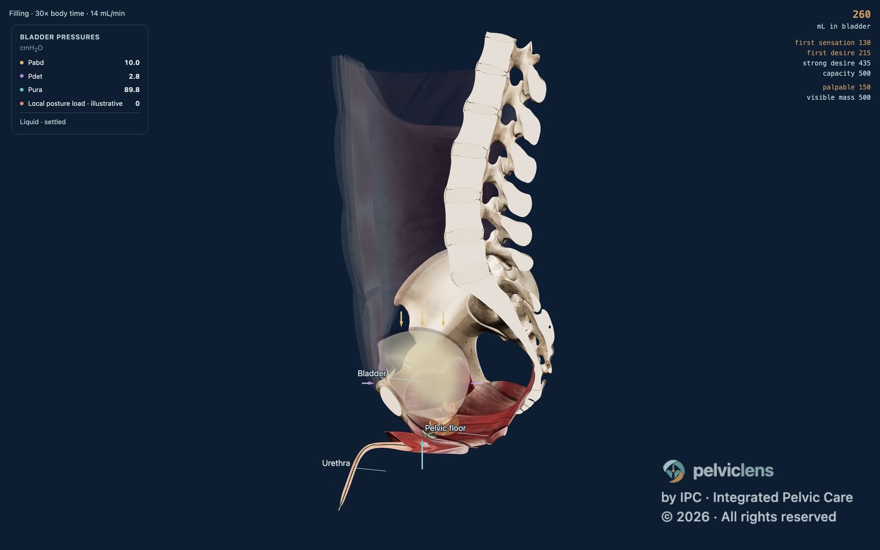 CystometrogramCystometrogram for the pelvic health clinician: the filling curve read as pressure, Pves = Pabd + Pdet, and what to tell your patient.17 min read
 Intrinsic sphincter deficiencyIntrinsic sphincter deficiency for the pelvic health clinician: why one valve is left after prostatectomy, and what to train first.16 min read
Read this article in Français · Español · Português
This article is education for health professionals. It is not medical advice, not a diagnosis and not a treatment plan for any person. Clinical decisions rest on your own assessment of the patient in front of you. If you are a patient reading this, talk it through with your own health professional. Evidence changes: the review date above tells you when this article was last checked.